Advanced 3D & 4D Ultrasound for Women’s Health
Obstetric 3D & 4D Ultrasound



See Your Baby. Understand Their Development. Cherish the Experience.
3D and 4D ultrasound can provide detailed images of your baby’s facial features, movements and development. When clinically appropriate, it complements routine pregnancy ultrasound by offering additional visual information while creating a meaningful bonding experience for parents.
Detailed views of your baby’s face and features
Real-time visualization of fetal movement
Assessment of selected fetal structures
Monitoring growth and development
A memorable bonding experience for parents
Gynaecological 3D Ultrasound
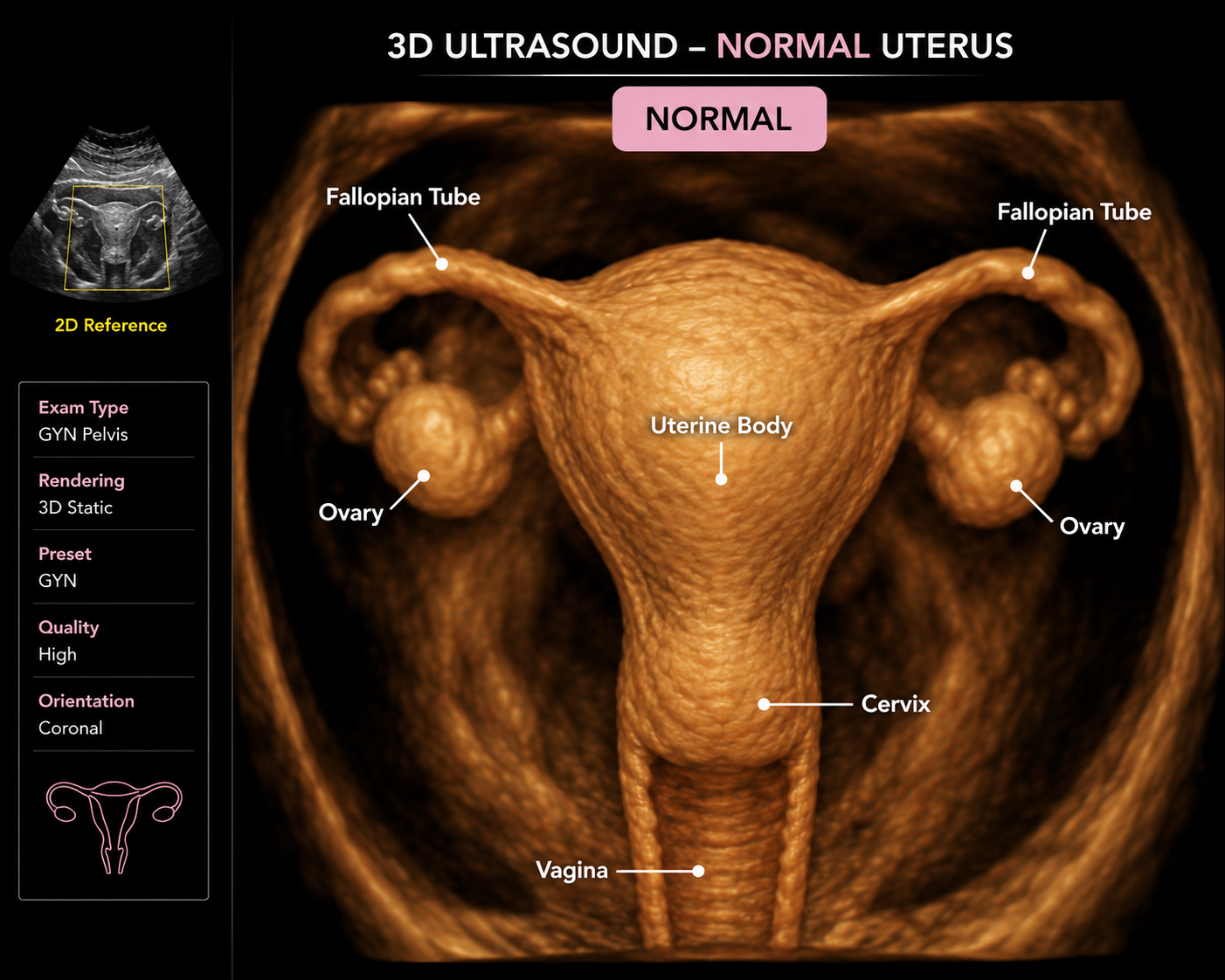
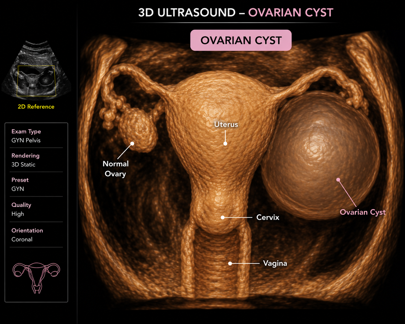
Clearer Imaging. Greater Diagnostic Confidence.
Advanced 3D gynaecological ultrasound provides detailed views of the uterus, uterine cavity and surrounding pelvic structures. It can help identify abnormalities that may not be clearly demonstrated with conventional two-dimensional imaging.
Uterine fibroids and polyps
Congenital uterine abnormalities
Adenomyosis
Ovarian and pelvic masses
Abnormal uterine bleeding
Assessment of the uterine cavity
Position of intrauterine contraceptive devices
Preoperative planning where appropriate
Urogynaecological 3D & 4D Pelvic Floor Ultrasound
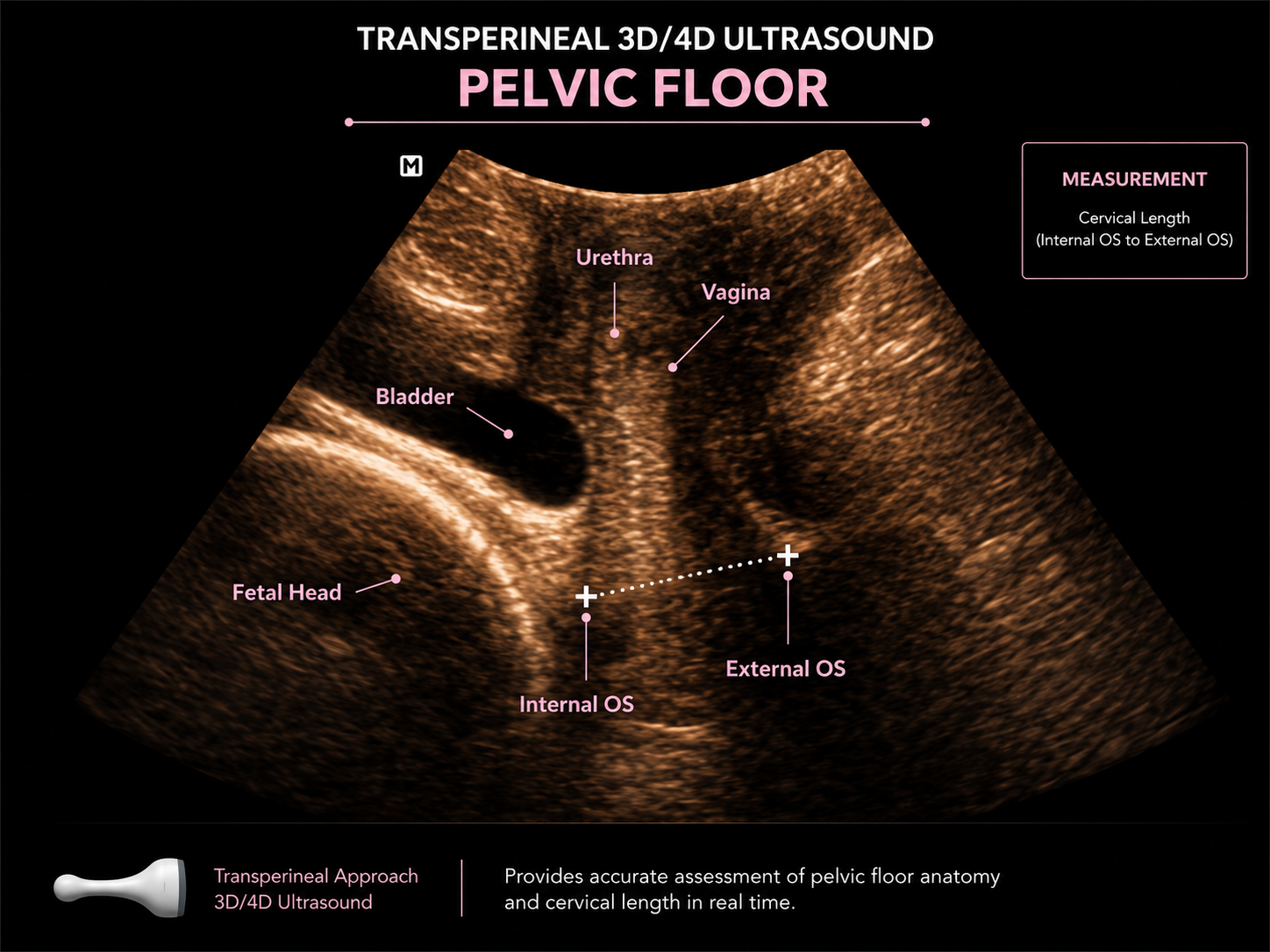
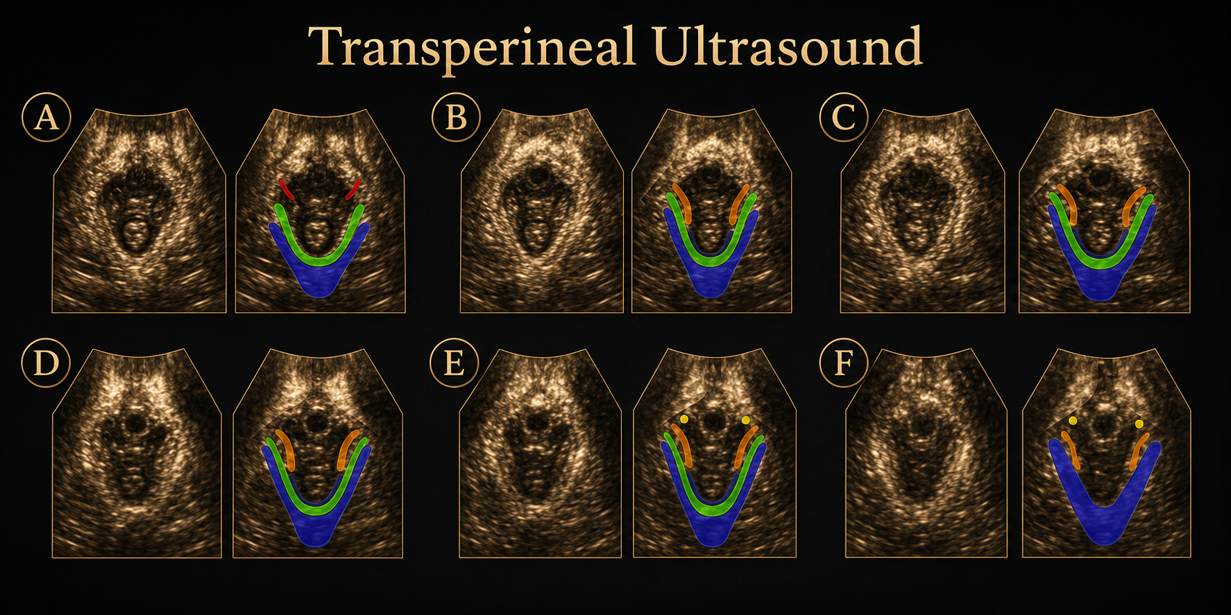
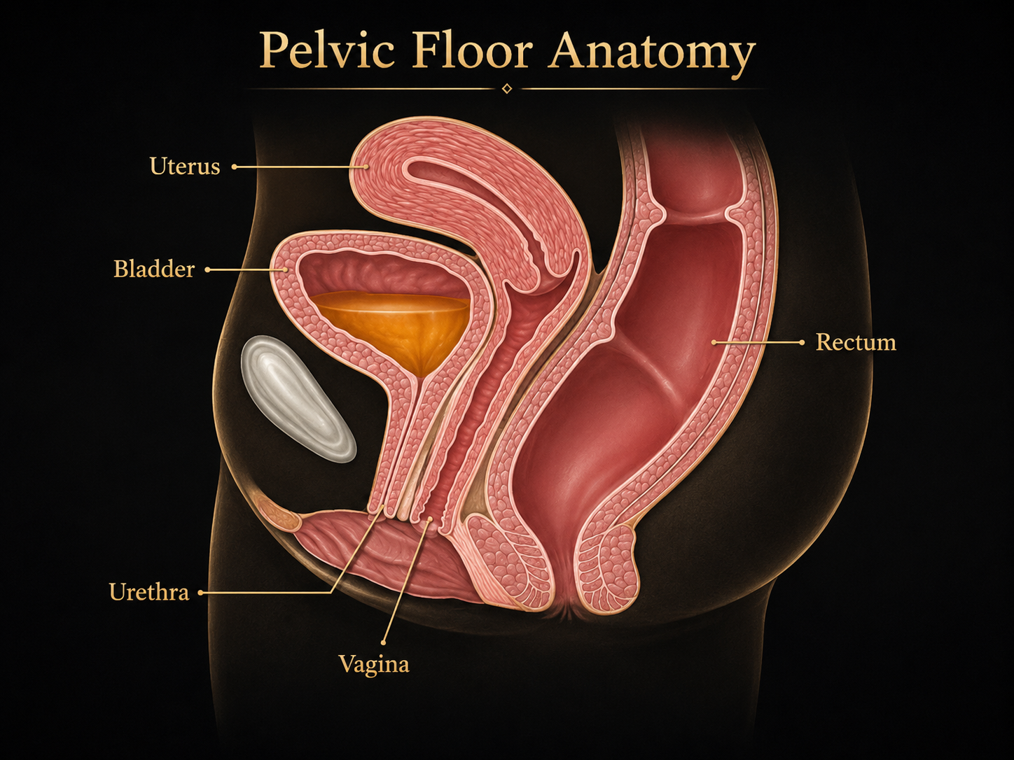
See the Pelvic Floor in Motion. Diagnose More Precisely.
Dynamic 3D and 4D transperineal ultrasound allows the pelvic floor to be assessed at rest, during contraction and while straining. It provides valuable information about how the bladder, vagina, pelvic organs and pelvic floor muscles move and function.
Pelvic organ prolapse
Bladder neck mobility
Stress urinary incontinence
Levator muscle injury following childbirth
Pelvic floor muscle function
Vaginal sling position
Mesh-related concerns
Complex or recurrent pelvic floor symptoms